
Heart rate variability (HRV) has been established as a non-invasive tool to study cardiac autonomic activity. Reduced HRV has been established as a predictor for increased risk of cardiac mortality and sudden cardiac death (1-6) especially in patients after myocardial infarction.
Several relaxation techniques have been established in the management of patients during cardiac rehabilitation aiming to reduce future cardiac events via cardiac autonomic nervous activity. It has been hypothesized that relaxation training programs may improve the cardiac autonomic nervous tone. However, this has so far not been proven for all available relaxation techniques, such as yoga.
Since more than 60 years B.K.S. Iyengar has been working therapeutically with patients after myocardial infarction. His method offers more than other techniques for relaxation do: the sequence of yoga asanas (exercises) and the individuality they are performed with, are adjusted to the severity of myocardial infarction, the stage of recovery and the clinical condition of the patient. Props are used, so that the strain of exercise can be regulated or stopped if desired.
We investigated whether this method has a positive influence on the cardiac vagal modulation compared to a similar setting of conventional relaxation and exercise techniques among healthy yoga practitioners.
Methods
Subjects
We examined 11 healthy yoga practitioners (7 women and 4 men, mean age: 43 ± 11; age range: 26-58 years). They were experienced practitioners with more than 3 years of regular practice of Iyengar-Yoga, four of them were certified teachers of Iyengar Yoga.
Each individual was subjected to 5 training units each of 90 minutes at the same time of the day (around 12.00 – 13.30 p.m.) once a week over 5 successive weeks. During two sessions they practiced Iyengar-Yoga; on three sessions they practiced a placebo program of relaxation that consisted of resting on the floor and park walking.
To avoid misinterpretations of our findings due to interindividual and circadian variability (7), the yoga practitioners themselves served as an intraindividual control group.
The group of yoga practitioners was also compared to an age and gender matched group of healthy individuals without evidence of cardiovascular disease who have not been practicing any relaxation techniques (n=11), to identify long-term effects.
All volunteers gave informed consent for scientific use of their Holter information.
Intervention
Before the actual examination started, the yoga practitioners had 3-5 sessions where they practiced the sequence of yoga asanas chosen for the intervention to make them familiar with the program. Since the yoga practitioners were experienced, the program, which was taken from the work of B.K.S. Iyengar addressed patients after myocardial infarction already at an advanced stage of recovery.
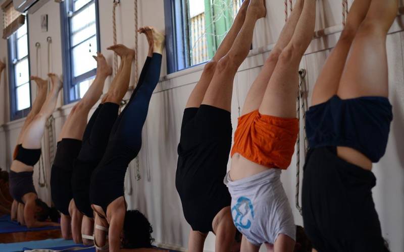
The program started with about 15 minutes resting poses, continued by 60 minutes standing poses, backbends and inverted poses and ended with another 15 minutes of resting poses. The sequence of asanas is shown in table 1.

Usually a training program for cardiac patients ends with the asana Bhismacharyasana; here we changed the series to end the program with Shavasana (lying on the floor). By this change we created three comparable blocks of body position between yoga and placebo program (see figure 1). Asanas were conducted by a certified teacher for Iyengar Yoga (K.K.).
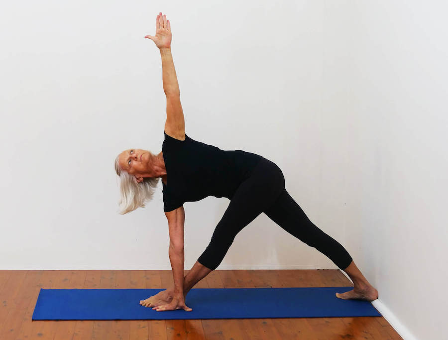
All asanas of B.K.S. Iyengar’s work with cardiac patients after myocardial infarction focus on opening the chest, therefore in standing poses a trestle was used and in supine position a special support for the thoracic spine was applied (e.g. wooden plank and small heart brick, figure 2+3). The exact description of the exercises is beyond the scope of this article. Details have been described by B.K.S. Iyengar elsewhere (8-9).
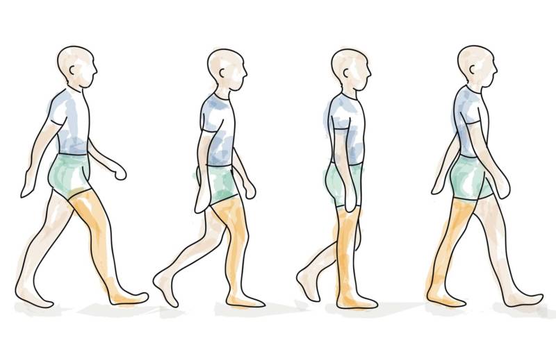
The placebo program was designed to be comparable to the yoga intervention and consisted of about 15 minutes resting on the floor in supine position, 60 minutes park-walking and again 15 minutes of resting on the floor. During park-walking and resting the trainees were also under guidance of the yoga instructor, and were taught to relax certain muscle groups.

Data Collection
During 5 successive weeks, once a week all volunteers underwent 24- hour ambulatory ECG monitoring with two-channel time-tracking Holter recorders (Tracker II, Reynolds, Herford, UK). The Holter Recording was initiated between 11 a.m. and 12 p.m. At around 12 p.m. the intervention program started. During 3 sessions they underwent the placebo program, during 2 sessions the yoga program was conducted.
All Holter recordings were manually edited by an experienced physician (H.B.) for exclusion of artifacts and premature beats. The physician was unaware of the group and the intervention. One R-R interval before and 5 R-R- intervals after an atrial or ventricular premature beat were eliminated from the analysis.
A minimum of 22 hours of analyzable data and a minimum of 90% of analyzable NN intervals were required for a tape to be accepted as valid. The median duration of the recordings was 24 hours, with 96% of valid analyzable NN intervals. There was no drop out.
Analysis of HRV
For each subject, time-domain HRV was measured according to the Task Force of ESC and NASPE (2) using a Pathfinder digital analysis system (Delmar Reynolds).
Mean RR interval and the following HRV parameters were calculated as hourly values and as 24- hour values: square root of the mean of the sum of the squares of differences between adjacent NN intervals (rMSSD), standard deviation of NN intervals (SDNN), mean standard deviation of NN intervals for all 5-minute segments (SDNNi), standard deviation of the averages of NN- intervals for all 5-minute segments (SDANN), absolute count of adjacent successive NN intervals differing by > 50 msec per hour (sNN50), and geometrical triangular index (TI).
Statistical Analysis
Statistical analyses were conducted with a commercially available software package (SPSS version 12.0; SPSS Inc). Comparisons between groups were performed utilizing a Mann-Whitney U test. Multiple comparisons were done by Bonferroni corrected analysis of variance for repeated measures. Consecutively an alpha corrected paired Student’s t test was performed for interval-to-interval comparisons. HRV parameters were tested for normal distribution with the Komolgorov-Smirnov goodness-of-fit test for normality. All parameters but sNN50 were normally distributed. Although natural logarithmic transformation could diminish the skewness of the distributions of sNN50, data was not transformed with regard to the lack of comparability with previous published data. HRV-data are presented as mean values ±standard deviation. Statistical significance was set up at p<0.05.
Results
The baseline criteria of yoga practitioners and the matched control group are shown in table 2.

There were no significant differences regarding hourly mean values of RR interval and parameters of HRV outside the intervention time.
Mean RR interval was significantly higher during the time of yoga intervention compared to placebo and to control (865 ±119 ms; 746±86 ms; 753±115 ms respectively, p<0.001for both).
The increase in the parameters of HRV was significantly higher during yoga exercise than during the placebo program and control especially for the parameters associated to the vagal tone [mean SD of NN intervals for all 5-minute intervals (SDNNi) 86.9 ± 16 vs 62.9± 53.3±18 (p< 0.001 for both); root mean square successive difference rMSSD 37.3 ±10 vs 30.1± 9 vs 24.1 ± 12 (p<0.01 for both)].
Estimates of overall HRV were significantly higher regarding the geometrical Triangular Index (TI) during yoga compared to placebo and control at time of intervention (26.5 ± 6 vs 24.6 ± 8 vs17.6 ± 6; p < 0.001 for both). SD of NN interval (SDNN) was not significantly different during yoga exercise compared to the placebo program, but was significant for both compared to the control at time of intervention (129.6 ± 22 vs 130.7± 32 vs 78.7 ± 26¸p < 0.001).
SD of the averages of NN intervals for all 5min segments (SDANN) – a long term parameter associated with physical activity- was higher during the placebo program of resting and parkwalking compared to the yoga program (90.8±32 vs 116.6± 33.5; p< 0.01) and significantly higher for both yoga and placebo compared to the control (p< 0.001) (figure 3 and table 3).


A different analysis compared mean HRV parameters of day and night with the time of intervention. Especially the vagus associated parameters (SDNNi and rMSSD) showed a typical nocturnal arousal as a sign of higher parasympathetic activity during the night. Only the increase during yoga intervention could compare to the peak at nighttime for these parameters (see fig. 4 and table 4).
Discussion
This study demonstrates that relaxation by yoga training is associated with a significant increase of cardiac vagal modulation among healthy yoga practitioners.
Since this method is easy to apply with no side effects, and leads to a deep physical and mental relaxation, it could be a suitable intervention during cardiac rehabilitation to shift the autonomic balance towards an increase of vagal activity and possibly decrease cardiac mortality.
La Rovere et al showed that exercise training by bicycle ergometry, an established training method in cardiac rehabilitation programs, increases vagal activity in patients after myocardial infarction (10), investigating baroreflex sensitivity as an autonomic marker. However, they reported that exercise training alone does not seem to be the only determinant of improved survival. It was only the combination of exercise with an increase in baroreflex sensitivity that predicted better survival. From experiments with dogs, Billmann et al considered the possibility that even independently from physical training, increased baroreflex sensitivity would be associated with a reduced risk for cardiac mortality after myocardial infarction (11).

Also in experiments with dogs after healed myocardial infarction, Kukielka et al (12) reported that submaximal long duration exercise reduced cardial vagal regulation initially, but further exercise training attenuated the initially exercise induced reductions in heart rate variability, suggesting a maintained higher cardiac vagal activity during exercise in the trained state.
On the other hand, Duru et al (13) found no significant effect of high-intensity exercise training on HRV indexes among patients with new-onset left ventricular dysfunction after myocardial infarction after 1, 2 and 12 months of training in a rehabilitation center, despite beneficial effects on clinical variables.
These findings suggest, that after myocardial infarction with resulting left ventricular dysfunction which makes up the majority of post-infarction patients, exercise training remains of limited value on HRV improvements. Furthermore, it seems that in a previously untrained condition, the subject needs first to develop a trained state, before exercise training can have a positive influence on cardial vagal regulation, and that during the phase of building-up exercise capacity an unwanted counter-effect might occur.

There is a risk that especially young and untrained patients tend to go beyond their (cardiac) exercise capacity. We also experience patients having reduced left ventricular function or comorbidities, who cannot tolerate regular exercise on bicycle ergometry.
In these cases, training by relaxation programs could be a suitable alternative and are already established in many rehabilitation centers as an additional training.
A systematic meta-analysis about relaxation therapy for rehabilitation and prevention in ischaemic heart disease by Dixhoorn et al of 27 controlled trials in which patients with myocardial infarction were taught relaxation therapy revealed that intense supervised relaxation practice enhances recovery from an ischaemic cardiac event (14). This meta-analysis included relaxation techniques such as progressive muscle relaxation, autogenic training, biofeedback, breath relaxation, hypnosis and psychological training.
Among these 27 controlled trials 3 studies investigated and revealed a positive effect on HRV (15-17). The applied techniques of relaxation were progressive muscle relaxation, breath relaxation, deep breathing, cue controlled relaxation and biofeedback
In our study we could show, that training after the method of B.K.S. Iyengar among healthy yoga practitioners was superior to a simple relaxation program that consisted of resting on the floor and mild exercise like park-walking. We think that his method might be superior to other relaxation techniques since it is a unique combination of relaxation (achieved by components like muscle stretching and relaxing, deep breathing, awareness [comparable to biofeedback], psychological aspects, concentration and meditation) and very exact therapeutical physical work that can be tailored for any limiting condition or co-morbidity.
The postures for cardiac patients are chosen and modified in a way to improve the loading – unloading conditions of the heart, which could positively influence remodeling and healing. In every asana the chest is kept open to improve respiration and achieve a higher oxygenation of blood. Back bending actions give a lengthwise stretch to the mediastinum.
Depending on stage of recovery and condition of the patient, the body is gradually brought to more inverted postures which increase venous return to the heart.
During yoga exercises the trainees had a lower heart rate than during the alternative program. Even during postures that build up body tension like standing poses or backbends, using slow and more isometric muscle contraction, the heart rate did not rise much.
A slow heart beat prolongs the diastolic filling of the heart, decreases myocardial oxygen consumption and increases myocardial perfusion.
A study among 24.913 patients by Diaz et al clearly identified a high resting heart rate (which also reflects cardial autonomic imbalance towards sympathetic activity) in patients with suspected or proved coronary artery disease as an independent predictor for total and cardiovascular mortality (18). Furthermore, a recently published study introducing deceleration capacity of heart rate – a novel Holter-ECG based marker for vagal activity – underlines the crucial role of cardiac vagal modulation regarding cardiovascular mortality in postinfarction patients undergoing modern treatment, particularly treatment involving acute revascularization procedures (19).
Further studies are required to investigate whether the demonstrated positive effect of therapeutical yoga on the cardiac vagal modulation can be transferred to cardiac patients and introduced into cardiac rehabilitation programs.
Study Limitations
The study cohort (regular yoga practitioners) warrants drawing conclusions about long lasting effects of yoga on HRV parameters. However, the 24 hour circadian rhythm of the yoga practitioners e.g. for SDNNi and rMSSD was higher than in the control group, yet the study population was too small to show a significant difference.
Additional studies are required to investigate long term effects of yoga training on cardiac autonomic nervous modulation.
Acknowledgement
We like to thank Mr. B.K.S. Iyengar for his guidance during this study. He tailored the sequence of asanas for this investigation and gave us a glimpse of his knowledge and experience of therapeutic yoga.
The original article has been published by the journal ecam (evidence based complementary and alternative medicine) in December 2007 (Khattab K, Khattab AA, Ortak J, Richardt G, Bonnemeier H. Iyengar yoga increases cardiac parasympathetic nervous modulation among healthy yoga practitioners. Evid Based Complement Alternat Med. 2007 Dec; 4:511-7.